
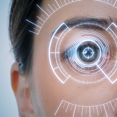
One does not see with their eyes, but with the help of the eyes, wherefrom the information is transmitted through the optic nerve, decussation, visual tracts to the certain areas of the occipital lobes of the cerebral cortex, where the picture of the world that we see, is formed. All these organs make up our visual analyzer or visual system.
Having two eyes makes our vision stereoscopic (forms a three-dimensional image). The right side of the retina of each eye transmits the "right side" of the image to the right side of the brain via the optic nerve, the left side of the retina acts similarly. Then two parts of the image – right and left – are connected together by the brain.
Since each eye perceives "its own" picture, when the joint movement of the right and left eye is disturbed, binocular vision may be impaired. It means, that you will start to have double vision or you will see two completely different pictures at the same time.

The eye can be called a complex optical device. Its main task – to "transmit" the correct image to the optic nerve. Eye structures perform different functions:
- an optical system, that projects the image;
- a system, that perceives and "encodes" the received information for the brain;
- a system, which "maintains" life support.
Cornea – transparent shell, that covers the front of the eye. There are no blood vessels in it, it has great refractive power. It is part of the optical system of the eye. The cornea borders the opaque outer shell of the eye – sclera.
Front camera – this is the space between the cornea and the iris. It is filled with intraocular fluid.
Iris – shaped like a circle with a hole inside (pupil). The iris consists of muscles, during contraction and relaxation of which the size of the pupil changes. It is part of the choroid of the eye. The iris is responsible for eye color (if it is blue – it means, it has few pigment cells, if it is brown – it means it has many pigment cells). It performs the same function, as the aperture in a photocamera, adjusting the light flow.
Pupil – a hole in the iris . Its dimensions usually depend on the level of illumination. The more light, the smaller the pupil.
The lens – "natural lens" of the eye. It is transparent, elastic – can change its shape, almost instantly "focusing", due to which a person can see well near, and far. Located in a capsule, it is held by a cilia strip. The lens, as well as the cornea, is part of the optical system of the eye.
Vitreous body – gel-like transparent substance, located in the back of the eye. The vitreous body supports the shape of the eyeball, participates in intraocular metabolism. It is part of the optical system of the eye.
Retina – consists of photoreceptors (they are sensitive to light) and nerve cells. Receptor cells, located in the retina, are divided into two types: cones and sticks. In these cells, that produce the enzyme rhodopsin, light energy is transformed (photons) into electrical energy of nervous tissue, meaning it is a photochemical reaction.
The sticks have high light sensitivity and allow you to see in poor lighting conditions, they are also responsible for peripheral vision. Cones, vice versa, require more light for their work, but they are the ones that allow you to see small details (are responsible for central vision), make it possible to distinguish colors. The largest concentration of cones is in the central fossa (macules), which is responsible for the highest visual acuity. The retina is adjacent to the choroid, but not too thoroughly in many places. It is here that it tends to detach in case of various diseases of the retina.
Sclera – opaque outer shell of the eyeball, which transforms in the front part of the eyeball into the transparent cornea. There are 6 oculomotor muscles attached to the sclera. It contains a small number of nerve endings and blood vessels.
Vascular membrane – lines the back part of the sclera, is adjacent to the retina, which it is closely related with . The choroid is responsible for the blood supply of intraocular structures. In case of the diseases of the retina, it is often involved in the pathological process. There are no nerve endings in the choroid, therefore, there is case of its desease, which in other cases can usually signal about certain visual disturbances.
Optic nerve – with the help of the optic nerve, signals from the nerve endings are transmitted to the brain.